Part Two
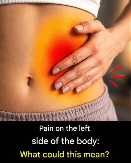
Left upper quadrant (LUQ) discomfort frequently implicates gastric mucosa, splenic capsule tension, or splenic flexure syndrome.
Splenic enlargement or subclinical micro-infarction stretches capsular innervation, producing a deep somatic ache.
Splenic flexure syndrome traps colonic gas at the sharp anatomical turn beneath the diaphragm, radiating shoulder or chest pressure.
Left renal pathology—such as acute pyelonephritis, renal calculi, or ureteral obstruction—presents as classic costovertebral angle tenderness.
Ureteral colic involves smooth muscle spasm responding to luminal crystal transit, triggering autonomic nausea and diaphoresis.
Left lower quadrant (LLQ) pain strongly prioritizes diverticulitis, sigmoid colon inflammation, or micro-perforation in susceptible demographics.
Diverticular outpouchings trap fecaliths, fostering localized bacterial proliferation and pericolic inflammatory phlegmon.
Gynecological considerations in biological females include left-sided ovarian torsion, ruptured functional cysts, or ectopic gestation.
Ovarian torsion cuts off vascular pedicle perfusion, causing acute, severe ischemic pain requiring surgical emergency evaluation.
Lower gastrointestinal etiologies also span infectious colitis, inflammatory bowel disease flares, or irritable bowel hypersensitivity.
Vascular emergencies like descending aortic aneurysm expansion or acute mesenteric ischemia present atypically with tearing deep left or central pain.
Referred visceral pain pathways from pancreatic tail pathology or lower left pleural bases can mimic abdominal wall or deep organ distress.
Diagnostic workup begins with thorough point-of-care history: radiation patterns, fever curves, bowel habit alterations, and urinary symptoms.
Laboratory evaluation inspects complete blood counts for leukocytosis, inflammatory markers like CRP, renal panel azotemia, and urinalysis for hematuria.
Cross-sectional imaging via contrast-enhanced CT of the abdomen and pelvis remains the radiological gold standard for acute quadrant evaluation.
Ultrasound safely assesses gynecological adnexal structures, free pelvic fluid, and renal hydronephrosis without ionizing radiation exposure.
Red-flag clinical markers mandating immediate emergency department triage include hemodynamic instability, rigid peritoneal guarding, high fever, or hematochezia.
Conservative outpatient management for mild non-complicated functional or dietary flare-ups involves temporary low-residue modification, hydration, and targeted antispasmodics.
Long-term recurrence prevention relies on dietary fiber optimization, visceral hypersensitivity modulation, and structured blood pressure/renal metabolic surveillance.
Concluding summary: Left-sided body pain is a multi-system diagnostic puzzle spanning gastrointestinal, urological, vascular, and gynecological domains; respecting anatomical geometry and red-flag thresholds transforms silent anxiety into precision clinical action.